Thanks Jong, this is indeed a very smart way of using random illumination ! Here is the paper:Hi Igor,.... I would like to bring your attention to our new paper which appears in Scientic Reports published by Nature.Fluorescent microscopy beyond diffraction limits using speckle illumination and joint support recovery at http://www.nature.com/srep/2013/130625/srep02075/full/srep02075.htmlAs you can see from the title, this work exploits the joint sparsity of the fluorescent probe distributions to improve the optical resolution beyond the diffraction limits. The main idea is to use random illuminations that modulate the emission intensities of flurophores whose locations do not changes during the illuminations. This makes the imaging problem a typical MMV (multiple measurement vector) problem, which can be solved using compressed sensing MMV algorithms. As you can see from the paper, using the standard fluorescent probes and proteins, we can improve the resolution by factor of 3. This is a big advantage in real experiments, since we do not need specially designed fluorescent probes as in STORM/PALM.I would be happy to hear any feedback from you !Best regards,-Jong
Fluorescent microscopy beyond diffraction limits using speckle illumination and joint support recovery by Junhong Min, Jaeduck Jang, Dongmin Keum, Seung-Wook Ryu, Chulhee Choi, Ki-Hun Jeong & Jong Chul Ye
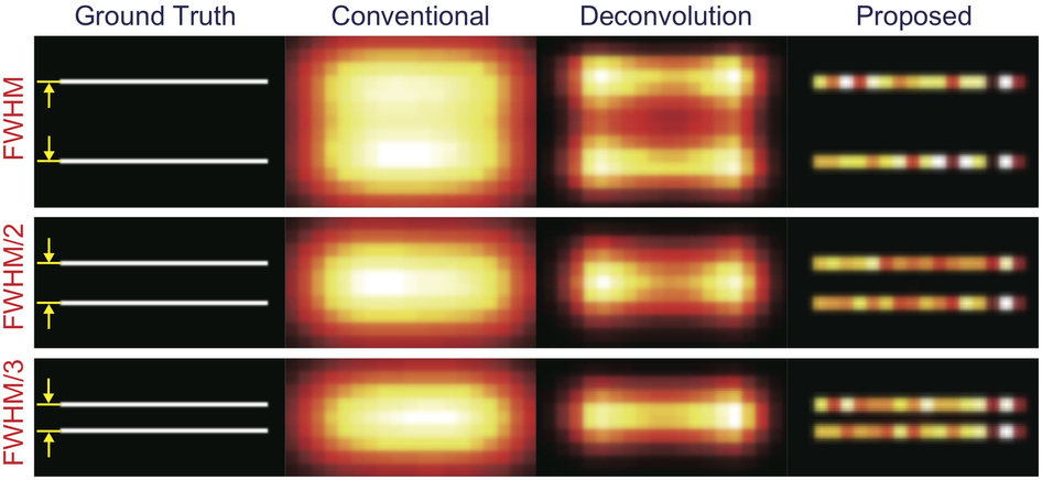
Structured illumination microscopy (SIM) breaks the optical diffraction limit by illuminating a sample with a series of line-patterned light. Recently, in order to alleviate the requirement of precise knowledge of illumination patterns, structured illumination microscopy techniques using speckle patterns have been proposed. However, these methods require stringent assumptions of the speckle statistics: for example, speckle patterns should be nearly incoherent or their temporal average should be roughly homogeneous. Here, we present a novel speckle illumination microscopy technique that overcomes the diffraction limit by exploiting the minimal requirement that is common for all the existing super-resolution microscopy, i.e. that the fluorophore locations do not vary during the acquisition time. Using numerical and real experiments, we demonstrate that the proposed method can improve the resolution up to threefold. Because our proposed method succeeds for standard fluorescence probes and experimental protocols, it can be applied in routine biological experiments.
Join the CompressiveSensing subreddit or the Google+ Community and post there !
 Liked this entry ? subscribe to Nuit Blanche's feed, there's more where that came from. You can also subscribe to Nuit Blanche by Email, explore the Big Picture in Compressive Sensing or the Matrix Factorization Jungle and join the conversations on compressive sensing, advanced matrix factorization and calibration issues on Linkedin.
No comments:
Post a Comment